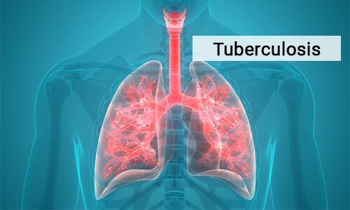
TUBERCULOSIS(TB)
DEFINITION
Tuberculosis (TB)
is an infectious disease caused by several species of mycobacterium that affects the lungs.
It is caused mainly by mycobacterium tuberculosis and rarely by mycobacterium bovis or mycobacterium africanum. Tuberculosis affects the lungs but may spread to almost any part of the body including the meninges, kidneys and bones.
TYPES OF
TUBERCULOSIS
Tuberculosis is classified as;
- Pulmonary tuberculosis, which affects the lungs
- Extra pulmonary tuberculosis, which affects any part of the body other than lungs.
PULMONARY TUBERCULOSIS
This is the most common cause of tuberculosis infection. It accounts for more than 80% of all cases of tuberculosis.
The type of pulmonary tuberculosis is as follows;
- Sputum smear – positive pulmonary tuberculosis (sm+PTB), this is a patient with sputum in which mycobacterium have been found on microscopy.
- Sputum smear – Negative pulmonary patient tuberculosis (MS –PTB).
This is a patient with sputum smear negative for
mycobacterium on microscopy, but x-ray shows consistence with active
tuberculosis which does not clear with ordinary antibiotics.
In some cases, even though sputum smear are all
negative for mycobacterium on microscopy, the culture is positive for
mycobacterium tuberculosis.
EXTRA PULMONARY
TUBERCULOSIS
This is a type of tuberculosis that affect any other
organ apart from the lungs. Types of
extra pulmonary tuberculosis are as follows;
1. Pleural
tuberculosis.
2. Glandular
tuberculosis(the glands)
3. Tuberculosis
of the spine
4. Tuberculosis
meningitis (the brain)
5. Intestinal
tuberculosis (intestines)
6. Miliary
tuberculosis (the whole body)
7. Tuberculosis
of the bone
8. Tuberculosis
of the skin
9. Tuberculosis
of the eye.
INCIDENCE
Tuberculosis can occur in persons of all ages but
especially young adults in the prime of life and the aged. Incidence is also high in the following people:
1. People
with acquired immune deficiency syndrome.
2. Babies
and young children who have weak immune systems.
3. People
who have been previously infected.
4. People
in close contact with someone who has infectious tuberculosis.
5. People
who consume alcoholic beverages and smoke heavily.
6. Incidence
is also high among health workers example nurses and doctors.
7. Mine
workers and people working in quarries
AETIOLOGY
Pulmonary tuberculosis results from exposure to
mycobacterium and sometimes other status of mycobacterium from a person with
active pulmonary disease who expels the organism through talking, coughing,
sneezing or singing or also from droplets in the air.
Mycobacterium bovis causes bovine tuberculosis and
this commonly affect cows or oxes.
Mycobacterium africancum is at variant of mycobacterium tuberculosis
both of which affect human beings.
PATHOPHYSIOLOGY
When a person inhales aerosol particles contaminated
by the germs of tuberculosis, they are carried into the lungs and into the
delicate epithelia surface of the alveoles, they cause swelling and local
capillary dilation.
Although some organisms will be engulfed by alveolar
macrophages, all will not be destroyed and will continue to multiply. The invasion of the lung tissue by the
bacteria give rise to an inflammatory reaction in which the affected alveoli
are filled with fluid, macrophages and bacteria. The degeneration of the lung tissue gives
rise to or forms a cheesy mass which is referred to as cessation. The resultant
lung damage eventually leads to fibrosis which will be visible on x-ray.
The primary lesion in the lungs is often asymptomatic
and confined to one area, and these tiny areas where the healing occurs by scar
formation and even by the deposit of calcium are called granulomas. A primary
infection may heal with the host acquiring immunity in the process, in some
cases, the primary lesion progresses to produce extensive disease locally or the
infection disseminate to produce metastic or miliary lesions. Thus some of the
bacteria will escape and may enter the blood stream to infect other organs of
the body. This form of tuberculosis is
called extra pulmonary or miliary tuberculosis.
Militry tuberculosis usually occurs in the lymph
nodes, meninges, joints, peritonium,
genito- urinary tract and bowel.
Tuberculosis present a chronic cough with mucopurulent
sputum as the disease becomes advanced. Dominant bacillinary also become
reactive when any of the predisposing factors or combination of them prevail.
SIGNS AND SYMPTONS
OF PULMONARY TUBERCLUSIS
·
Clinical
Manifestation of pulmonary tuberculosis include;
1. A
bad cough that lasts longer than two weeks
2. Pain
in the chest
3. Coughing
up blood or sputum ( phlegm from deep inside the lungs)
4. Shortness of breath
·
General
signs and symptoms of tuberculosis
1. Dry
persistent cough
2. Weakness
and fatigue
3. Profuse
night sweats
4. Mucopurulent
or blood stained sputum
5. Haemoptysis
6. Dyspnoea
with chest pains
7. Weight
loss
8. Low
grade fever with intermittent temperature
9. Anorexia
10. On
auscultation, crepitant cranks, bronchial breath sounds and wheezing may be
heard.
11. Pallor
and anaemia.
12. There
is low grade fever.
COMPLICATIONS
Pleural effusion
Hoarseness of voice
DIAGNOSTIC
INVESTIGATIONS
Specific
Diagnostic Test
i)
Sputum smear test –
Gastric aspiration, laryngeal swab for ziehl- Nelson stains and fluorescent
microscopic examination for tuberculosis and also for culture and sensitivity
to antibiotics. Positive sputum indicates open tuberculosis
ii)
Radiology of chest and
lungs show the shadows of inflammation and there may be calcification
especially in the upper lobes of the lungs.
iii)
Positive tuberculin skin
test
iv)
sputum smear for AFB
(Acid fast Bacilli)
v)
Purified protein
derivatives (PPD)
vi)
Screen test or multiple
puncture test
vii)
Chest X-ray
MEDICAL TREATMENT
·
DRUG
TREATMENT
The
national tuberculosis programme (NTP) uses three types of standardised treatment
regimen. All regimen start with an initial intensive phase followed by a
continuation phase.
(A)
THE
SHORT COURSE
This course is for;
1. New
smear positive pulmonary tuberculosis patient.
2. Patients
whose sputum smears are negative but who are seriously ill.
The course consists of two months intensive phase
followed by six months continuation phase. The dosage given usually depends on
the age and weight of the patient.
The drugs used for the intensive phase are:
i)
Streptomycin (intramuscularly)
Dosage:
Adult – 1g daily for two months (reduces dosage for elderly and emaciated)
Children – 20mg1kg body weight daily for
2months.
ii)
Rifiriah (intramuscularly
or orally)
Dosage:
Adult – 1g daily for two months.
Children: 10mg1kg body weight for two months.
iii)
Pyrazinamide (orally)
Dosage:
Adult – 2g daily for two months
Children – 35mg1kg body weight for two months.
The drug for the continuation phase is;
·
Thiacetazone (orally)
Adult
dosage: 450mg daily for six months.
Children
·
Rifiriah ( orally or
intramuscularly)
Dosage:
10mg1kg body weight daily for six months
If there is a high suspicion of HIV infection in a
patient, the thiacetazone may be change to isoniazid plus ethambutol
·
Ethambutol (orally)
Dosage
400mg daily for six months
·
Isoniazid ( orally or
intramuscularly)
Dosage 300mg daily for
six months.
(B)
STANDARD
COURSE
This consists of 12months duration for smear negative
pulmonary tuberculosis and extra pulmonary tuberculosis cases.
It comprises of two months intensive phase treatment
with streptomycin, isoniazid and thiacetazone.
It is followed by 10months continuation phase with
isoniazid and thiacetazone.
Administration of thiacetazone can cause Stevens
Johnson syndrome in patients with HIV hence are not put on standard course.
(C) RETREATMENT COURSE
This is for relapse and treatment failure cases.
It comprises of initial intensive phase of five drugs
·
Rifampicin
·
Isoniazid
·
Pyrazinamide
·
Ethambutol.
Daily
for three months supplemented with streptomycin for two months. This phase is
strictly supervised and if possible the patient is admitted.


Comments
Post a Comment